Home
Future Meetings
How to Join
Past Meetings
SMSI Awards
Publications
History
Contacts

SMSI Awards 2005
Emile Chamot Award Recipient

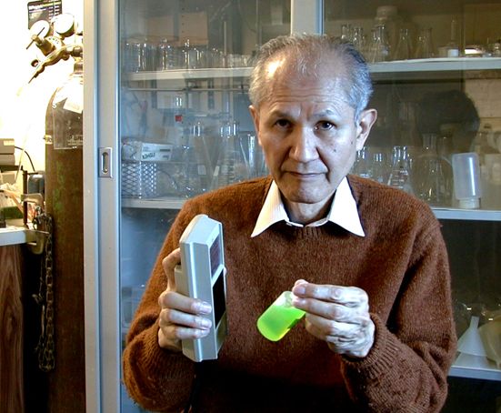
Dr. Osamu Shimomura for contributions to Fluorescence Microscopy
by isolating aequorin and green fluorescent protein
How aequorin and green fluorescent protein were discovered
We discovered aequorin and the green fluorescent protein (GFP) in 1961 from the luminous jellyfish Aequorea. They are unusual proteins; the former emits blue light in the presence of a trace of calcium ions even in the absence of oxygen, and the latter is brilliantly green fluorescent even in day light. By the end of 1970s, we were able to characterize most of the important properties of the two proteins including the chemical structures of their functional chromophores. Helped by the progress in genetic research, both proteins were cloned, apoaequorin in 1985 and GFP in 1992, making it possible to generate them even in live cells. Now both proteins are indispensable research tools, aequorin as a calcium probe and GFP as a marker protein. In retrospect, however, it looks as if my Aequorea project had been programmed by my three mentors for its success. In 1955, a professor of the Nagasaki Pharmacy College, for whom I was working as a teaching assistant, kindly allowed me to do research at the Hirata lab at Nagoya University. Professor Hirata assigned me to do the study of Cypridina luciferin, which eventually gave me the knowledge that is essential to solve the problems of Aequorea. Then, Dr. Frank Johnson invited me to his lab at Princeton University in 1960, and he gave me the subject of Aequorea to study. The guidances given to me were indeed in exact order for solving the difficult problems of aequorin and GFP.
| History | 1967-1979 | 1980-1999 | 2000 | 2001-2004 | 2005 | 2006-2011 | 2012 | 2013 | 2014 | 2015 | 2016 | 2017 | 2018 | 2019 |